Biopsies
US/MRI Fused Trans-Perineal Biopsies
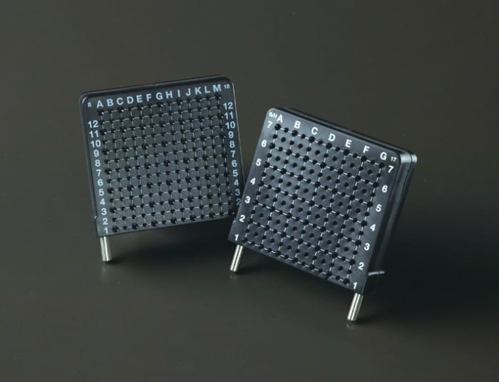
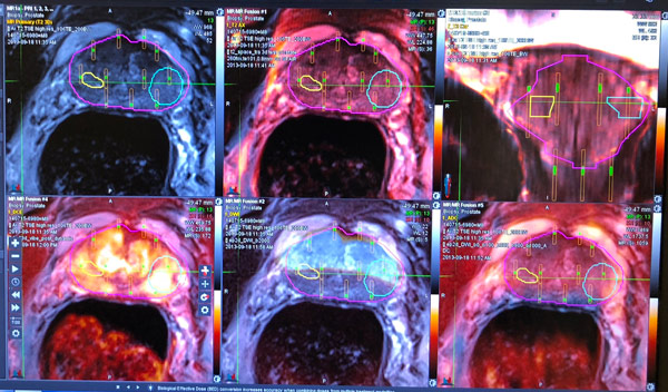
Traditional prostate transrectal biopsies are by far the most frequently used method to diagnose prostate cancer. All biopsy procedures use an interstitial perineal template for guidance, not free-hand, as most others not trained in template guidance technique use. At Desert Prostate Specialists, we have significantly advanced the prostate biopsy procedure to a trans-perineal, not trans-rectal, method using MRI/Ultrasound fusion and 3D reconstruction to achieve a safer, painless and more comprehensive procedure. How Does it Work? Under general outpatient anesthesia, in the surgery center, an ultrasound transducer is inserted into the rectum. Images are obtained and fused with the patient’s MRI scan in real time. Then, a template is attached to the transducer to use as a guide for the biopsy needle. This needle is then inserted through the template between the scrotum and rectum and into the perineal skin to access the prostate gland samples. This is all done under computer guidance and visualized on a large screen TV in the operating room where 3D reconstruction of the prostate occurs.

Why Use a Template for Guidance?
Template guidance has been the mainstay of interstitial techniques in the radiation oncology community for decades. It has a proven track record of allowing precision placement of needles for biopsy or therapy. Free hand techniques, as used by others, are not able to provide the guidance necessary for the precision placement of needles into the areas of risk as defined by the ultrasound/MRI fusion techniques. Guidance means precision, and precision means accurate and reproducible results. If the biopsy is positive, these techniques allow for precision and guided treatments for better cure rates and less toxicity. At Desert Prostate Specialists, our excellent results and low toxicity risks speak for themselves.

How do Patients Benefit?
Because the procedure is performed under general anesthesia, the procedure is painless. As no needle is inserted into the rectum and the biopsy is performed in a surgery center, the procedure is safe with very little risk of bleeding or infection. We are able to obtain biopsies from regions of the prostate gland that are only accessible through the trans-perineal technique. Finally, because the procedure uses MRI/Ultrasound fusion and there is genetic testing follow-up of benign results, this type of biopsy is far more comprehensive. We get the answers we need about your condition.
